|
|
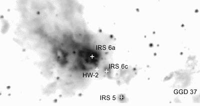
A grayscale image of Cep A in the K' band showing the infrared reflection nebula, the location of GGD37, the compact radio source HW-2 and the infrared "peaks" reported by Lenzen et al. 1984. North is up and east is to the left. The field of view is approximately 3' x 1.5'. |
|
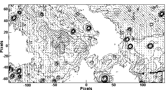
J image of Cep A. The 0,0 position corresponds to HW-2. The image scale is 180 x 90 arcsecs with a plate scale of 0.675 arcsec/pixel. North is up and east is to the left. Polarization vectors are superimposed after binning 4 x 4 pixels for clarity; 100% polarization is equivalent to 10 arcseconds. A total of 48 minutes of integration time was used in this image which 45 seconds of on-chip integration time represented by the contours. There is a small region of missing data in the lower right corner due to an imperfect mosaic. |
|
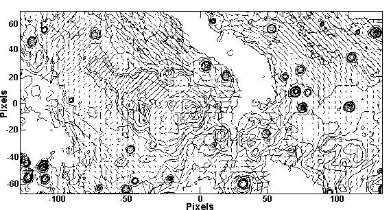
H image of Cep A, same attributes as Figure 1. A total of 28 minutes of observing time was used with 15 or 20 second on-chip integrations. There is a small region of missing data in the upper right corner due to an imperfect mosaic. |
|
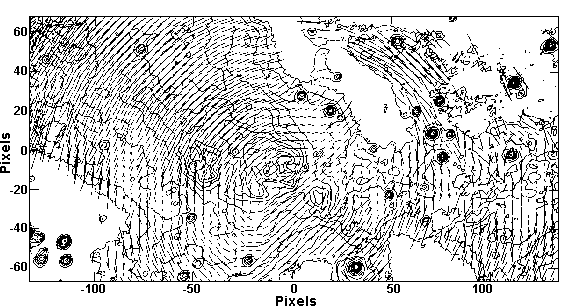
K' image of Cep A, same as for Figure 1. A total of 76 minutes of integration time is shown with the flux levels representing 6 seconds on-chip. There is no missing data in this mosaic. Note the reduced extinction compared to Figure 1. |